The common belief that 20/20 vision equals perfect vision is a myth; your brain’s ability to process dynamic information is far more critical for real-world performance.
- Standard eye exams test static acuity (what you see), but ignore dynamic processing (how you react to what you see).
- Two distinct neural pathways—the « what » and « where/how » systems—govern these functions, and an imbalance can cause issues despite clear eyesight.
Recommendation: If you have « perfect » acuity but struggle with visual fatigue, slow reactions, or motion sensitivity, it’s time to investigate your functional vision with a specialist.
You’ve just left the optometrist’s office, certificate in hand: 20/20 vision. You can read the smallest letters on the chart from 20 feet away. By all standard measures, your eyesight is perfect. Yet, something feels off. You struggle to track a fast-moving ball in a weekend sports game, feel a wave of nausea as a passenger in a car, or experience a deep, draining fatigue after an hour of driving at night. You’re told your eyes are fine, but your daily experience tells a different story. This frustrating paradox is not in your head; it’s in your brain.
The fundamental misunderstanding stems from equating visual acuity—the clarity of your eyesight measured by a static eye chart—with the entirety of your visual ability. This is like judging a high-performance computer solely on its screen resolution while ignoring its processing speed. Your eyes are the cameras, but your brain is the dual-core processor responsible for making sense of the visual world. More specifically, it runs two distinct « operating systems » in parallel: a system for identifying *what* you see, and a completely separate one for processing *where* it is and *how* to interact with it.
The 20/20 test only evaluates the « what » system. It completely overlooks the « where/how » system, which governs your reaction time, spatial awareness, and ability to process motion. This article will dismantle the myth of 20/20 perfection by exploring the science of these dual visual pathways. We will explain why you can have flawless acuity but still struggle with dynamic tasks, how to assess these hidden skills, and what it means to train your brain, not just your eyes, for truly superior visual performance.
This guide breaks down the complex world of visual processing into understandable concepts. We will explore the different facets of your vision, explain why standard tests fall short, and introduce the specialists who can truly diagnose and treat these functional challenges.
Summary: Decoding the Truth Behind Perfect Vision and Brain Processing
- Why Can You Read the Bottom Line But Still Struggle With Moving Objects?
- How to Assess Your Visual Reaction Time for Gaming or Driving?
- High Contrast Sensitivity or High Acuity: Which Is More Important for Night Driving?
- The Static Chart Trap: Why Standard Exams Miss Dynamic Vision Issues?
- When to Train Your Peripheral Awareness to Reduce Motion Sickness?
- Why Is the Cover Test Essential for Detecting Lazy Eye?
- Why Does Peripheral Vision Improvement Boost Reaction Time in Team Sports?
- What Does an Orthoptist Do That an Optician Cannot Treat?
Why Can You Read the Bottom Line But Still Struggle With Moving Objects?
The answer lies in the brain’s division of labor. Neuroscientists have identified two primary visual pathways that process information from your eyes, often called the « Two-Streams Hypothesis. » The first is the ventral stream, or the « what » pathway. This system travels to the temporal lobe and is responsible for object recognition, detail analysis, and color perception. It’s the part of your brain that aces the eye chart by identifying those tiny, static letters. When you have 20/20 acuity, your ventral stream is performing flawlessly.
The second, and often overlooked, pathway is the dorsal stream. This is your « where/how » system. It sends information to the parietal lobe and is responsible for processing spatial awareness, motion, depth perception, and guiding your physical actions in response to what you see. It tells you where an object is, how fast it’s moving, and how to coordinate your body to catch, hit, or avoid it. As neuroscientists explain, this pathway is the key to interaction. As Multiple researchers note in the well-established Two-streams hypothesis:
The dorsal stream is in charge of spatial awareness and guidance of actions (e.g., reaching).
– Multiple researchers, Two-streams hypothesis – Wikipedia
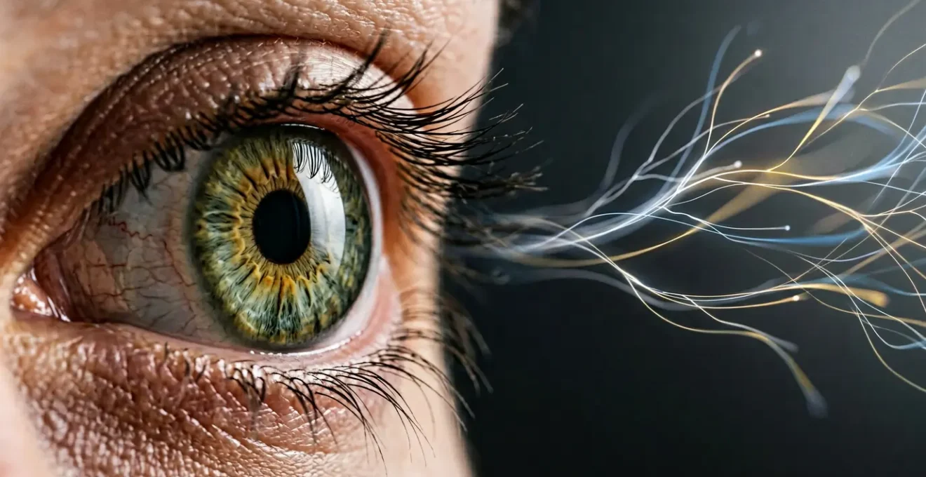
This functional split is why you can have a « high-resolution camera » (great acuity) but a « slow processor » for motion. Your brain is getting a crystal-clear image from the eyes, but the system responsible for interpreting movement and location is lagging. The struggle isn’t with *seeing* the moving object; it’s with *processing* its trajectory and speed in real-time.
As the visual above suggests, static recognition and dynamic interaction are handled by entirely different systems. A standard eye exam that only tests your ability to identify stationary targets completely fails to assess the efficiency of your crucial « where/how » pathway, leaving the root cause of your struggles with moving objects completely undiagnosed.
How to Assess Your Visual Reaction Time for Gaming or Driving?
While a formal evaluation requires a specialist, you can begin to gauge your dynamic visual skills with simple tools and observations. Visual reaction time is not just how fast you click a mouse; it’s the entire sequence from detecting a stimulus to initiating a physical response. This process is a core function of the dorsal stream. Elite performers in visually demanding fields like esports offer a clear benchmark; research shows that pro-level competitive gamers achieve reaction times of 150-180 ms, significantly faster than the average person.
Online reaction time tests can provide a baseline. These tests measure your « simple reaction time » to a single, anticipated stimulus (e.g., a screen changing color). However, real-world scenarios like driving or gaming involve « choice reaction time, » where you must respond differently to various stimuli. For a more functional self-assessment, pay attention to your performance in these environments. In a video game, do you consistently notice an opponent in your periphery and react before they get the drop on you? When driving, how quickly do you notice a car braking several vehicles ahead and adjust your speed accordingly?
These are all tests of your dynamic visual processing. A lag in these situations, despite clear vision, points to an inefficiency in your « where/how » system. Observing these real-world delays is often the first step in recognizing that your visual performance goes far beyond the static letters on an eye chart. To get a more structured sense of where you stand, you can perform a simple audit.
Your 5-Point Dynamic Vision Self-Audit
- Peripheral Awareness Test: While focusing on a point directly in front of you, have a friend move their fingers in your far peripheral vision. Note how far out you can detect the motion without moving your eyes.
- Saccadic Eye Movement: Rapidly switch your focus between two objects placed several feet apart. Do your eyes jump smoothly and accurately, or do you find yourself over- or under-shooting the target?
- Depth Perception Check: Try pouring water into a narrow glass. Do you do it effortlessly, or do you often misjudge and spill? This simple task relies heavily on accurate binocular depth cues.
- Visual Tracking: Follow a fast-moving object with your eyes, like a tennis ball during a match. Can you keep your eyes locked on it, or do you lose track and have to find it again?
- Choice Reaction in Practice: Use an online tool that requires you to respond differently to different colors or shapes. This better simulates the decision-making aspect of real-world dynamic vision than a simple reaction test.
High Contrast Sensitivity or High Acuity: Which Is More Important for Night Driving?
When driving at dusk or in the dark, the world is not a high-contrast eye chart. It’s a palette of grays, shadows, and low-light objects. In this environment, your ability to distinguish an object from its background—a skill known as contrast sensitivity—is far more critical than your ability to read a small sign. High acuity (20/20 vision) means you can see a small, black letter on a bright white background. High contrast sensitivity means you can spot a deer standing on the gray shoulder of an unlit road.
Research consistently shows that contrast sensitivity is a better predictor of night driving performance than visual acuity. For instance, a 2022 study found that mesopic visual acuity and disability glare index accounted for 21% of variation in hazard detection times among older drivers, highlighting that vision under low-light conditions is a complex skill. Your eye’s photoreceptors, the rods and cones, must adapt to the dark, and your brain must work harder to interpret the limited information it receives. This is a processing challenge, not an acuity one.

The U.S. Federal Highway Administration has reached a definitive conclusion on this matter, directly linking this functional skill to safety. Their findings leave no room for ambiguity about what truly matters on the road after sunset. As they state in their research summary:
Contrast sensitivity is noted as being more impactful on vehicular crashes than is visual acuity.
– Federal Highway Administration, FHWA Roadway Visibility Research
If you have 20/20 vision but feel anxious or unsafe driving at night, it’s highly likely you have reduced contrast sensitivity. You may be technically *seeing* clearly, but your brain isn’t receiving enough data to effectively distinguish shapes and objects in the low-light, low-contrast environment. This is yet another clear example of how functional vision, not static acuity, dictates real-world performance and safety.
The Static Chart Trap: Why Standard Exams Miss Dynamic Vision Issues?
The standard eye exam is designed to do one thing very well: measure refractive error and test static visual acuity. It determines if you are nearsighted, farsighted, or have astigmatism, and whether corrective lenses can bring your « what » pathway to the 20/20 standard. This is the « Static Chart Trap. » Because it is so effective at its narrow purpose, it has created the false impression that it is a comprehensive assessment of vision. It is not.
Standard exams do not typically test for:
- Eye Teaming (Binocularity): How well your two eyes work together as a synchronized team.
- Eye Focusing (Accommodation): How quickly and accurately your eyes can shift focus from near to far objects.
- Eye Tracking (Saccades): How smoothly your eyes can follow a moving object or jump between targets.
- Contrast Sensitivity: Your ability to detect objects in low-contrast environments.
- Peripheral Awareness: How well you notice and process information at the edges of your visual field.
These are all functional vision skills managed primarily by the dorsal « where/how » stream. Diagnosing and treating deficits in these areas requires a completely different approach, known as a Functional or Developmental Vision Evaluation. This assessment goes beyond the static chart to measure how your eyes and brain work together in dynamic, real-world situations. The cost for such a specialized assessment reflects its comprehensive nature, with comprehensive functional vision testing typically ranging from $250 to $600. It’s an investment in understanding your true visual capabilities.
Performing these tests requires not only specialized equipment but also advanced training that most standard optometrists do not possess. As experts in the field explain, it’s a sub-specialty.
Only optometrists who have undergone advanced post-graduate training in the field of vision development, and who have the necessary equipment required to test for and treat functional vision problems, can perform this detailed assessment of visual function.
– Texas Vision Therapy, Functional Vision Evaluations
This is why millions of people can pass a standard vision screening with flying colors yet continue to suffer from undiagnosed functional vision problems that cause headaches, eye strain, and poor performance in sports or driving.
When to Train Your Peripheral Awareness to Reduce Motion Sickness?
Motion sickness, or kinetosis, occurs when there is a conflict between what your eyes see and what your inner ear’s vestibular system senses. When you’re a passenger in a car, your inner ear feels the motion, but if you’re looking at a static phone screen, your central vision tells your brain you’re stationary. This sensory mismatch triggers feelings of nausea, dizziness, and discomfort. The key to mitigating this often lies in your peripheral vision.
Your peripheral vision is highly attuned to detecting motion—it’s a primary function of the dorsal « where/how » stream. By looking out the side window of a moving vehicle, you allow your peripheral vision to see the landscape flowing past. This visual input of motion aligns with what your vestibular system is feeling, resolving the sensory conflict and often reducing or eliminating the symptoms of motion sickness. Therefore, you should actively engage your peripheral vision whenever you are in a situation that is known to trigger your motion sickness.
Scientific research supports this connection between the dorsal stream and the perception of self-motion (known as vection). In one key experiment, researchers used brain imaging to see how the brain processed visually-induced self-motion. The results were revealing.
Case Study: The Brain’s Motion Sickness Hub
A 2012 study published in *BMC Neuroscience* used PET scans to observe brain activity during circular vection (the feeling of spinning induced by watching a rotating visual field). The study found that the *intensity* of the perceived motion was processed in the parietal areas of the brain—the heartland of the dorsal stream. This directly demonstrates that the « where/how » pathway, which heavily relies on peripheral input, is the system responsible for processing the very sensory information that can lead to motion sickness. Training this system to better handle peripheral motion can therefore be a direct therapeutic approach.
If you frequently suffer from motion sickness, it can be a strong indicator that your brain is inefficient at processing peripheral visual motion. Instead of avoiding motion, a more effective long-term strategy can be to actively, but gradually, train your peripheral awareness. Simple exercises, like practicing focusing on a central point while paying attention to objects moving in your periphery, can help your brain become more comfortable and efficient at integrating this type of sensory input, ultimately reducing your susceptibility to motion sickness.
Why Is the Cover Test Essential for Detecting Lazy Eye?
Lazy eye, or amblyopia, is a perfect example of a developmental vision problem that has nothing to do with acuity in the affected eye itself. It is a brain problem, not an eye problem. Amblyopia occurs when the brain, during early childhood, learns to ignore or suppress the information coming from one eye. This often happens due to a misalignment of the eyes (strabismus) or a significant difference in refractive error between them. The eye itself may be perfectly healthy, but the brain simply doesn’t « listen » to it.
The cover test is the primary diagnostic tool for detecting this issue because it directly assesses binocular function—how the two eyes work together as a team. During the test, the optometrist has the patient focus on a target while they cover one eye, then the other. The practitioner closely observes the uncovered eye for any movement. If the uncovered eye has to move to pick up fixation on the target, it indicates that it wasn’t properly aligned or working with the other eye to begin with. This simple, non-invasive test reveals a breakdown in the brain’s ability to fuse two separate images into a single, three-dimensional view.
This directly relates to our dual-stream model. Proper binocular vision is a foundational skill for the dorsal « where/how » stream. Without accurate input from both eyes, the brain cannot calculate depth perception or spatial relationships correctly. A person with untreated amblyopia might have 20/20 vision in their « good » eye, but they are functionally « blind » to 3D space. They will struggle with tasks like catching a ball, navigating stairs, or parking a car because their brain is not receiving the data needed to perform these spatial calculations.
Detecting and treating amblyopia early with tools like the cover test is crucial. Treatments such as vision therapy and patching work by retraining the brain to pay attention to the suppressed eye, re-establishing the neural connections necessary for proper binocular function. It underscores a core principle of neuro-optometry: you don’t see with your eyes; you see with your brain.
Key Takeaways
- 20/20 acuity only measures your static « what » system; it ignores the dynamic « where/how » system responsible for reaction and coordination.
- Your brain uses two distinct pathways (ventral for identification, dorsal for spatial interaction), and an imbalance between them causes visual issues.
- Functional vision skills like contrast sensitivity, peripheral awareness, and eye teaming are more critical for real-world tasks like night driving and sports than static acuity.
Why Does Peripheral Vision Improvement Boost Reaction Time in Team Sports?
In any fast-paced team sport, what happens at the edge of your vision is just as important as what happens directly in front of you. An athlete’s ability to perceive a teammate cutting to the basket, a defender approaching from the side, or the overall spacing of players on the field is what separates good players from great ones. This skill, known as peripheral awareness, is a hallmark of an elite dorsal stream. Because this « where/how » system is optimized for detecting motion, improving its efficiency directly translates to faster reaction times.
When you train your peripheral vision, you are essentially upgrading your brain’s « early warning system. » You learn to detect and process critical information without having to make a slow, conscious eye movement (a saccade) to look directly at it. This allows you to initiate a motor response—passing the ball, dodging a tackle—based on information processed pre-consciously. Experienced video gamers, who operate in a highly demanding visual environment, demonstrate this neurological advantage. Research shows that experienced gamers exhibit higher saccadic peak velocities, meaning their eyes physically move faster and more efficiently between targets.

This isn’t just theory; modern neuroscience has proven this link at a structural level. A groundbreaking study on video gamers provided definitive evidence that training these skills physically changes the brain.
Case Study: How Gaming Rewires the Brain for Faster Reactions
A 2023 study using functional MRI (fMRI) found that action video gamers have enhanced functional and structural connectivity within the dorsal visual stream. Specifically, they showed stronger connections between parts of the brain responsible for processing visual information and those responsible for spatial awareness. This heightened connectivity in the « where/how » pathway was directly correlated with faster response times in visual tasks, demonstrating that extensive practice fundamentally rewires the brain to be more efficient at turning peripheral visual input into rapid action.
For an athlete, this means that training peripheral awareness isn’t just an « eye exercise. » It is a form of neurological conditioning that builds a faster, more robust connection between seeing and doing. By enhancing the dorsal stream, you are literally speeding up the brain’s ability to react to the complex, dynamic environment of a team sport.
What Does an Orthoptist Do That an Optician Cannot Treat?
Understanding the different roles in eye care is crucial to getting the right help. While people often use the terms interchangeably, an optician, an optometrist, and a specialist like an orthoptist or developmental optometrist have vastly different scopes of practice. An optician is a technician trained to fit and dispense eyeglasses and contact lenses based on a prescription provided by an optometrist or ophthalmologist. They do not test vision or diagnose or treat eye conditions.
A standard optometrist is a primary eye care doctor who performs comprehensive eye exams to test for refractive errors (prescribing glasses/contacts) and to screen for eye diseases like glaucoma and cataracts. However, most do not specialize in the diagnosis and treatment of functional vision problems.
This is where the specialists come in. An orthoptist or a developmental optometrist is an expert in how the brain, nerves, and eye muscles work together. They are the professionals who diagnose and treat the very dorsal stream issues we’ve been discussing. Their focus is not on making you see the chart more clearly, but on making your visual system function more efficiently. As the experts at SeeVividly.com explain:
A Developmental Optometrist specializes in behavioral optometry which is concerned with how your eyes and visual system function and is interested in how your behavior affects vision or how your vision influences your behavior.
– SeeVividly.com, What is a Developmental Optometrist?
They treat conditions that an optician is completely unequipped to handle, and which a standard optometrist would typically refer out.
Case in Point: Post-Concussion Vision Syndrome
A patient who suffers a concussion may develop convergence insufficiency, a condition where the eyes are unable to work together to focus on a near object, leading to double vision, headaches, and dizziness. An optician cannot help. A standard optometrist will identify the lack of disease but may not offer treatment. A developmental optometrist or orthoptist, however, will perform a functional vision evaluation to diagnose the specific neuromuscular problem and prescribe a course of vision therapy—a series of exercises designed to retrain the brain-eye connection and restore proper function. This is a therapeutic intervention, not an optical one.
If you have 20/20 vision but still experience the frustrating symptoms of poor visual processing, the next logical step is to seek a comprehensive Functional Vision Evaluation from a qualified developmental optometrist or orthoptist. This is the only way to accurately diagnose and create a plan to treat the underlying cause of your difficulties.