The fundamental difference is not in treating the eyes, but in retraining the brain’s control system—a neurological function that corrective lenses cannot address.
- Orthoptists diagnose and manage failures in the neuromuscular control of the eyes, which manifest as double vision, eye strain, and reading difficulties.
- Unlike compensatory tools like glasses from an optician, orthoptic vision therapy offers a rehabilitative or curative pathway by leveraging neuroplasticity.
Recommendation: If visual symptoms persist despite an accurate glasses prescription, an orthoptic assessment is the critical next step to investigate the root cause.
You have the correct prescription from your optician, yet reading remains a taxing chore, leaving you with headaches. Or perhaps after a mild stroke or concussion, you experience a disorienting, intermittent double vision that glasses don’t fix. These scenarios highlight a common and frustrating gap in eye care—a gap that exists between the physical health of the eye and its functional performance. While an ophthalmologist manages ocular disease and an optician dispenses corrective lenses, a third, critical specialist operates in this functional space: the orthoptist.
The prevailing understanding often simplifies orthoptics to « eye exercises, » a trivialization that misses the core principle of our work. The issue is rarely a lack of muscle strength. The problem lies within the intricate neuromuscular feedback loop between the eyes and the brain. An orthoptist is a clinical specialist in the diagnosis and non-surgical management of this binocular vision system. Our role is to identify and treat the underlying processing errors that cause misalignment, poor focusing, and inefficient eye tracking.
But if the problem is in the brain’s control, how can it be fixed? The answer lies in neuroplasticity—the brain’s ability to reorganize itself by forming new neural connections. This is the foundation of orthoptic treatment. This article will deconstruct the specific functions of an orthoptist, distinguishing our diagnostic and therapeutic approach from any other optical profession. We will explore how we diagnose the root cause of misalignment, the mechanisms behind effective therapy, and when a referral is not just beneficial, but essential for reclaiming functional vision.
To understand this specialized field, this guide breaks down the core functions and treatment philosophies that define orthoptic practice. The following sections detail the diagnostic methods, therapeutic options, and critical timelines that differentiate our work from that of an optician.
Summary: The Orthoptist’s Role in Neurological Vision Rehabilitation
- Why Is the Cover Test Essential for Detecting Lazy Eye?
- How to Do Pencil Push-Ups Correctly to Fix Convergence Insufficiency?
- Prism Glasses or Vision Therapy: Which Solution Fixes Double Vision Permanently?
- The Consistency Trap: Why Stopping Therapy After 2 Weeks Reverses Progress?
- When to Ask for an Orthoptic Referral for Your Child’s Reading Issues?
- Why Does Your Latent Phoria Become Symptomatic After Age 40?
- Why Does Eye Alignment Affect Professional Confidence in Face-to-Face Roles?
- Sudden Double Vision: When Is It a Sign of a Binocular Disorder?
Why Is the Cover Test Essential for Detecting Lazy Eye?
The cover test is the cornerstone of orthoptic diagnosis, yet its purpose is widely misunderstood. It is not a simple check for acuity; it is a precise diagnostic procedure to unmask the brain’s underlying control of eye alignment. When a patient looks at a target, their brain works to fuse the images from both eyes into a single, three-dimensional picture. By covering one eye, we break this fusion. The true test is not what the covered eye does, but observing the uncovered eye’s movement as fusion is re-established. A subtle shift, jump, or drift reveals a phoria (a latent tendency to misalign) or a tropia (a manifest misalignment).
This objective measurement is critical in pediatric cases, especially for detecting amblyopia, or « lazy eye. » A child’s visual system is highly adaptable. The brain, faced with a misaligned eye, may choose to suppress or ignore its input to avoid double vision, leading to poor visual development in that eye. The cover test reveals this misalignment long before a child is old enough to complain or even notice a problem. It quantifies the neuromuscular deficit, providing the baseline for any therapeutic intervention. An optician cannot perform this diagnostic assessment.
To the untrained eye, the test seems simple. To the orthoptist, the speed, direction, and magnitude of the refixation movement provide a wealth of information about the patient’s fusional reserves and the nature of their binocular instability. It is a window into the brain’s automatic visual processing.
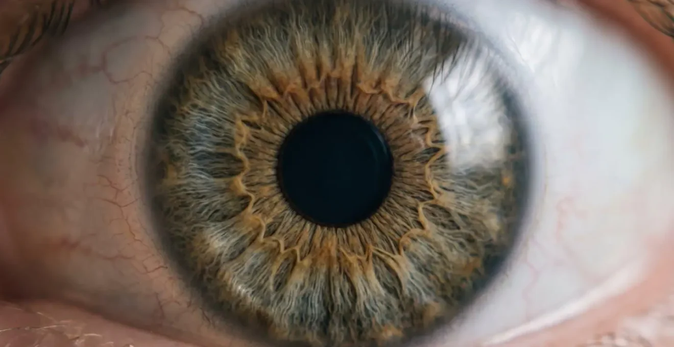
As this visualization of the diagnostic process shows, the technique allows us to observe the minute, involuntary movements that indicate how the brain is struggling to align the eyes. It is this objective data that separates a clinical assessment from a simple vision screening and forms the basis of a targeted therapy plan.
How to Do Pencil Push-Ups Correctly to Fix Convergence Insufficiency?
Pencil push-ups are perhaps the most well-known—and often incorrectly performed—orthoptic exercise. They are prescribed for convergence insufficiency (CI), a common binocular vision disorder where the eyes have difficulty turning inward together to focus on a near object. In fact, research demonstrates that up to 5% of school-age children experience CI, significantly impacting their ability to read comfortably. An optician might identify the symptom of near-vision strain but cannot prescribe the therapeutic protocol to correct the underlying neuromuscular cause.
The goal is not muscular strengthening; it is to improve neuromuscular control and sensory feedback. Correct execution is paramount. First, hold a pencil at arm’s length with a single, small letter as the target. Slowly bring the pencil towards your nose, concentrating on keeping the letter single and clear. The moment the letter doubles or becomes blurry, stop. This is your « break point. » The therapeutic task is not to push through the double vision, but to recognize this point and consciously try to regain a single image. Once single vision is restored, hold it for a few seconds before moving the pencil slightly closer. If you cannot regain a single image, move the pencil back out until it becomes single again.
The key is to avoid suppression. Many patients, in an effort to « succeed, » will unconsciously ignore the input from one eye, meaning they are seeing a single image but are not truly converging. An orthoptist teaches the patient to recognize the feeling of true binocular convergence versus suppression. This exercise, when done correctly, retrains the brain to maintain fusion at closer distances, reducing eye strain, headaches, and reading difficulties associated with CI. It is a foundational element of a much broader, office-based vision therapy program.
Prism Glasses or Vision Therapy: Which Solution Fixes Double Vision Permanently?
When a patient presents with diplopia (double vision) due to eye misalignment, there are two primary non-surgical pathways: compensatory prisms or rehabilitative vision therapy. An optician can dispense prism lenses, but only an orthoptist can diagnose the underlying cause and determine if a permanent, therapeutic solution is possible. The choice between these two approaches hinges on a critical distinction: are we compensating for the problem or curing it?
Prism glasses are a compensatory tool. They work by bending light before it enters the eye, effectively tricking the brain into perceiving a single image even though the eyes remain physically misaligned. This can provide immediate and significant symptom relief. However, it is a passive solution. The underlying neuromuscular deficit remains unchanged, and the patient becomes dependent on the glasses. In some cases, the brain may adapt to the prism, requiring progressively stronger prescriptions over time.
Vision therapy, on the other hand, is a curative approach. It is an active treatment program designed to retrain the brain to control eye alignment and movement correctly. Through a series of specialized activities and procedures, vision therapy leverages neuroplasticity to develop or restore the brain’s ability to fuse the images from both eyes naturally. As shown by a comparative analysis of treatment outcomes, this approach addresses the root cause of the problem.
| Treatment Approach | Mechanism | Permanence | Success Rate | Duration |
|---|---|---|---|---|
| Prism Glasses | Compensating: Bends light to help misaligned eyes | Temporary (requires ongoing use) | 75-100% immediate symptom relief | Instant relief |
| Vision Therapy | Curing: Retrains brain to control eye alignment | Permanent neuroplastic changes | 75-87% functional improvement | 4-8 months |
| Prism with Therapy | Combined: Temporary prism support during retraining | Progressive weaning to permanent cure | Higher success for large deviations | 6-12 months |
The decision is clinical. For some patients, such as those with paralytic strabismus or very large, stable deviations, prisms may be the only viable non-surgical option. But for many forms of binocular vision dysfunction, especially convergence insufficiency or decompensating phorias, vision therapy offers the potential for a permanent solution, eliminating the need for a lifelong visual crutch.
The Consistency Trap: Why Stopping Therapy After 2 Weeks Reverses Progress?
A common and dangerous misconception about vision therapy is that early, rapid improvement signals a cure. A patient may begin therapy, experience a dramatic reduction in symptoms within the first couple of weeks, and conclude the treatment is complete. This is the « consistency trap. » Stopping therapy prematurely almost guarantees a full regression of symptoms because it fundamentally misunderstands the science of neuroplasticity.
Initial progress in vision therapy is often the result of recruiting and strengthening existing, but underutilized, neural pathways. It feels like a quick fix because the brain is learning a new « trick. » However, this new skill is fragile. The old, dysfunctional habits of seeing are deeply ingrained over years or even decades. The real work of vision therapy is not just learning the new skill, but cementing it through repetition until it becomes the brain’s new, automatic default. This process of creating stable, permanent neural pathways requires sustained effort over time.
As the Convergence Insufficiency Treatment Trial (CITT), a landmark series of studies, demonstrated, true rehabilitation is a process. Follow-up data from these studies reinforce the necessity of completing a full therapeutic course and, in some cases, continuing with maintenance activities. As one analysis notes:
Patients who did not receive maintenance therapy showed higher rates of recurrence than those who maintained some level of continued visual activity.
– Convergence Insufficiency Treatment Trial (CITT), Follow-up data from CITT studies
This is why a full course of orthoptic therapy is not measured in days, but in weeks and months. The typical evidence-based program lasts between 12 and 24 weeks. This duration is not arbitrary; it is the time required to move beyond conscious effort and achieve automatic, reflexive control of the binocular system. Stopping early is like building a house and walking away after framing the walls—the structure is there, but it will not withstand the first storm.
When to Ask for an Orthoptic Referral for Your Child’s Reading Issues?
A child struggling with reading is often first evaluated for learning disabilities like dyslexia or attention disorders like ADHD. While these are valid considerations, a critical and frequently overlooked cause is binocular vision dysfunction (BVD). An optician ensures the child has 20/20 acuity, but clear sight is not the same as functional vision. Reading is one of the most demanding visual tasks, requiring the eyes to converge, focus, and track accurately for sustained periods. When this system fails, the child experiences symptoms that mimic other learning difficulties.
The statistics are telling: comprehensive studies reveal that up to 50% of children with reading problems may have an undiagnosed vision disorder. A parent should be alert to specific behavioral compensations. Does the child frequently lose their place on the page, skip words or lines, or use a finger to track? Do they complain of headaches, eye strain, or words looking « blurry » or « like they’re moving » after a short period of reading? A key physical sign is an abnormal posture, such as a persistent head tilt or holding reading material unusually close to their face, as the child instinctively tries to find a position that makes it easier for their eyes to work together.

These are not signs of laziness or a lack of concentration; they are hallmark indicators of a struggling binocular vision system. If a child has passed a standard vision screening (i.e., can read the eye chart) but exhibits these reading-specific behaviors, it is a clear signal to dig deeper. An optician’s role ends with providing clear glasses; an orthoptist’s work begins with investigating why a child with clear vision cannot read comfortably.
Your Action Plan: Key Signs to Discuss with a Specialist
- Behavioral Observations: Document specific reading behaviors (losing place, skipping lines, using a finger to track, short attention span for reading only).
- Symptom Reporting: Note any complaints of headaches, eye strain, blurry or double vision, or words moving on the page, especially after near work.
- Postural Compensations: Photograph or video any consistent head tilting, closing one eye, or holding books or screens abnormally close during reading.
- Performance Discrepancy: Compare reading ability and comprehension with verbal intelligence. Is the child bright and articulate but struggles significantly with text?
- Previous Screenings: Gather all past vision screening and optician reports. Even if « normal, » they provide a crucial baseline for the orthoptist.
Why Does Your Latent Phoria Become Symptomatic After Age 40?
Many adults are surprised by the sudden onset of double vision, eye strain, or headaches in their 40s and 50s, especially when they have never had vision problems before. This phenomenon is often the « decompensation » of a lifelong, latent phoria. A phoria is a small, natural misalignment of the eyes that the brain’s fusional reserves have been effortlessly controlling for decades. You have had it your whole life, but your brain’s robust compensatory mechanism has kept it asymptomatic.
The trigger for this sudden decompensation is almost always the onset of presbyopia, the age-related loss of focusing ability at near. The focusing system (accommodation) and the convergence system are neurologically linked. In youth, when you focus up close, you get a « free » boost of convergence. This accommodative convergence helps keep your latent phoria in check. After age 40, as the eye’s lens stiffens and your focusing ability weakens, this link breaks down. The accommodative system can no longer provide that extra boost, and your brain’s fusional reserves are suddenly forced to manage the entire misalignment on their own. After decades of relative ease, this new, constant demand quickly leads to fatigue and the breakdown of binocular control.
The result is the sudden appearance of symptoms from a condition you’ve unknowingly had your entire life. An optician will correctly diagnose the presbyopia and prescribe reading glasses, but this only solves the focusing problem. It does not address the now-symptomatic alignment problem. This is where an orthoptist is essential to diagnose the underlying phoria and prescribe targeted vision therapy to rebuild the depleted fusional reserves, restoring comfortable binocular vision.
Case Study: Adult-Onset Convergence Insufficiency
To illustrate this, consider the findings from a 20-year retrospective study on adult-onset convergence insufficiency. The study of 118 adults revealed a median diagnosis age of 68.5 years, demonstrating that the onset of presbyopia after 40 often serves as the trigger that weakens the accommodative convergence system. This weakening unmasks the previously compensated latent phoria. The mean exodeviation at near was 14.1 prism diopters, highlighting how a significant, previously controlled misalignment can suddenly become symptomatic as age-related accommodative flexibility is lost.
Why Does Eye Alignment Affect Professional Confidence in Face-to-Face Roles?
The impact of binocular vision dysfunction extends far beyond physical discomfort; it can profoundly erode professional confidence and performance, particularly in roles requiring significant face-to-face interaction. An individual with an intermittent tropia (a misalignment that appears under conditions of fatigue or stress) may find themselves struggling to maintain eye contact during meetings. To the individual, this is a fight to avoid seeing double. To colleagues, it can be misinterpreted as disinterest, shiftiness, or a lack of confidence.
The cognitive load required to maintain fusion can be immense. While colleagues are focused on the content of a presentation, the person with BVD is dedicating a significant portion of their mental energy to the physical act of seeing. This can lead to reduced comprehension, difficulty formulating responses, and overwhelming fatigue after client interactions or team meetings. The resulting headaches, dizziness, or even nausea can make a full day in the office feel like a marathon, impacting productivity and career advancement.
This struggle is often silent and invisible. The individual may not even connect their professional difficulties to their vision, attributing it instead to anxiety or burnout. An orthoptic diagnosis can be a moment of revelation, finally providing a physical explanation for what felt like a personal or psychological failing. As one patient described the impact of their untreated BVD:
A patient described becoming severely debilitated by binocular vision dysfunction, unable to drive for more than 10 minutes, shop for groceries, or manage basic housekeeping tasks. Nausea and dizziness limited daily activities significantly. Through visual rehabilitation, the patient reclaimed major aspects of life, regaining the ability to drive, work, and engage socially with confidence—demonstrating the profound impact of binocular disorders on professional and personal functioning.
– Patient success story
By restoring automatic, comfortable binocular vision, orthoptic therapy frees up the cognitive resources previously dedicated to the struggle of seeing. This not only eliminates physical symptoms but can restore the professional confidence needed to thrive in face-to-face environments. An optician can make you see clearly; an orthoptist can help you function confidently.
Key Takeaways
- Orthoptists are not « eye exercise » coaches; they are clinical specialists who treat the neurological control system governing eye alignment and movement.
- Unlike glasses from an optician which compensate for symptoms, orthoptic vision therapy aims to be curative by retraining the brain for permanent functional improvement.
- Persistent symptoms like headaches, reading difficulty, or intermittent double vision—especially if a correct glasses prescription is already worn—strongly warrant an orthoptic referral.
Sudden Double Vision: When Is It a Sign of a Binocular Disorder?
The sudden onset of double vision is an alarming symptom that requires immediate and careful differentiation. It can be the sign of a life-threatening neurological event, such as a stroke or brain tumor, or it can be the benign (though disruptive) decompensation of a pre-existing binocular vision disorder. As an orthoptist, a key part of our role is to help distinguish between these causes after urgent medical issues have been ruled out by a primary care physician or emergency department.
Emergency red flags that must prompt an immediate trip to the emergency room include double vision accompanied by a droopy eyelid, uneven pupils, facial numbness, severe headache, dizziness, or slurred speech. These are not symptoms of a simple binocular disorder and require urgent neurological evaluation. An orthoptist does not manage these acute emergencies.
However, double vision can also arise from less sinister causes. A common scenario is post-concussion syndrome, where the trauma has disrupted the brain’s ability to coordinate the eyes. Similarly, systemic illness, extreme fatigue, or the onset of presbyopia can overwhelm the brain’s capacity to manage a latent phoria, causing it to « break » into a manifest, double-seeing tropia. This is the « straw that broke the camel’s back »—a system that was just barely coping finally fails. In these cases, the double vision is often intermittent, worse when tired, and resolves when one eye is closed. While an optician is not equipped to manage this, an orthoptist is the correct specialist to diagnose the specific type of misalignment and begin a rehabilitative program to rebuild fusional control.
If you or a loved one experiences sudden double vision and all immediate neurological emergencies have been ruled out by a medical doctor, an orthoptic assessment is the critical next step. We provide the diagnosis and non-surgical treatment plan to restore single, comfortable binocular vision. If these symptoms resonate with your experience or that of your child, the next logical step is to seek a formal binocular vision assessment. Do not dismiss persistent visual discomfort; request a referral to an orthoptist to investigate the underlying cause and explore rehabilitative solutions.
Frequently Asked Questions about What Does an Orthoptist Do That an Optician Cannot Treat?
What are the emergency warning signs requiring immediate medical attention?
Seek emergency care immediately if double vision is accompanied by: droopy eyelid, uneven pupils, facial numbness or weakness, severe headache, dizziness, slurred speech, or signs of stroke. These indicate potential neurological emergencies requiring urgent evaluation.
What characterizes a benign decompensating phoria versus a neurological emergency?
Benign decompensation is typically intermittent, triggered by fatigue, illness, or new glasses, has a history of eye strain, and vision becomes single when one eye is closed. It develops gradually over hours or days rather than suddenly, and is not accompanied by neurological symptoms.
What causes a latent phoria to suddenly become symptomatic?
A lifetime of managing small latent eye misalignment can suddenly give way due to factors like age-related changes (especially after 40 with presbyopia onset), increased visual demands from prolonged digital device use, systemic illness, stress, or fatigue. This is the ‘straw that broke the camel’s back’ phenomenon where compensatory mechanisms finally fail.